
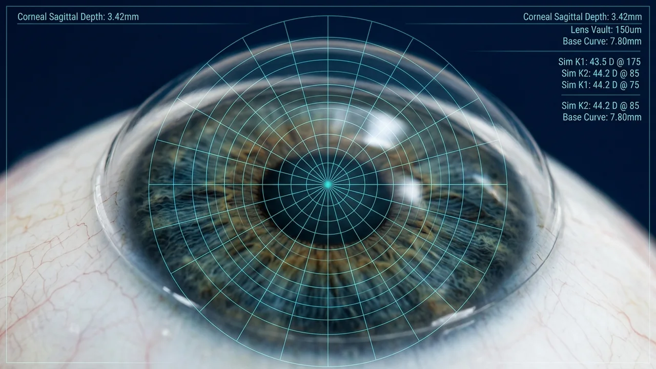
How a scleral lens sits on a keratoconic cornea
A scleral lens vaults the whole cornea and limbus and lands on the conjunctiva over the sclera. The fluid reservoir beneath it gives a smooth optical surface over the irregular cone.
- Contact lens
- Fluid reservoir
- Cornea with a cone
- Sclera
Schematic, not to scale.
Corneal ectatic disorders present significant challenges in achieving good visual quality. Among these conditions, keratoconus is one of the most common, characterized by progressive thinning and protrusion of the cornea, resulting in irregular astigmatism, myopia and increased higher-order aberrations.
While spectacles and conventional contact lenses may provide satisfactory vision in the early stages, progressive corneal irregularity can make conventional correction increasingly difficult. For these patients, scleral contact lenses have emerged as an important option for visual rehabilitation.
Understanding corneal ectasia
Corneal ectasia refers to a group of disorders characterized by progressive or structural thinning and distortion of the cornea. Important conditions include:
- Keratoconus
- Pellucid marginal degeneration (PMD)
- Post-LASIK and post-refractive surgery ectasia
- Post-keratoplasty irregular corneas
- Corneal irregularity following trauma or surgery
These conditions produce irregular astigmatism and higher-order aberrations, which can significantly affect visual acuity, contrast sensitivity and quality of life.
Early diagnosis, appropriate monitoring and timely referral for interventions such as corneal collagen cross-linking, when indicated, are essential components of comprehensive management.
What makes scleral lenses different?
Unlike conventional corneal rigid gas-permeable lenses, scleral lenses are larger-diameter gas-permeable lenses that vault over the entire cornea and limbus, resting on the conjunctival tissue overlying the sclera.
A fluid-filled reservoir is created between the posterior surface of the lens and the anterior cornea.
This unique design provides three important advantages:
1. Neutralization of corneal irregularity
The fluid reservoir creates a relatively smooth refractive surface, reducing the optical effects of irregular corneal curvature and higher-order aberrations.
2. Improved comfort and lens stability
Since the lens rests on the sclera rather than directly bearing on the sensitive corneal surface, many patients experience improved comfort and more stable vision compared with corneal RGP lenses.
3. Protection of the ocular surface
The fluid reservoir maintains a hydrated interface over the cornea and can be particularly beneficial in selected patients with ocular surface abnormalities.
Clinical applications in keratoconus
Scleral lenses are particularly valuable in patients with:
- Moderate to advanced keratoconus.
- Decentered or highly irregular corneal cones.
- Poor centration or instability with corneal RGP lenses.
- Intolerance to conventional RGP lenses.
- Significant irregular astigmatism following corneal procedures.
- Persistent visual symptoms despite spectacle correction.
Importantly, scleral lenses are not necessarily reserved for end-stage keratoconus. Depending on corneal shape, ocular surface characteristics and patient requirements, they can be considered at different stages of the disease.
Scleral lenses: a valuable option beyond keratoconus
The clinical applications of scleral lenses extend well beyond keratoconus.
They may offer significant benefits in:
- Pellucid marginal degeneration.
- Post-LASIK ectasia.
- Post-radial keratotomy (post-RK) irregularity.
- Post-penetrating keratoplasty.
- Corneal scarring and irregular astigmatism.
- Selected cases of Stevens-Johnson syndrome.
- Severe ocular surface disease and exposure-related keratopathy.
- Selected patients with complex anterior segment abnormalities.
Patient selection and fitting strategies must be individualized according to the underlying condition.
The fundamentals of successful scleral lens fitting
Successful scleral lens fitting involves more than simply selecting an appropriate diameter and base curve. A comprehensive assessment should include:
1. Pre-fitting evaluation
- Detailed ocular and contact lens history.
- Visual acuity and refraction.
- Slit-lamp examination.
- Corneal topography and tomography.
- Assessment of the conjunctiva and scleral contour.
- Evaluation of ocular surface health.
2. Corneal and limbal clearance
The lens should vault the cornea and limbus without compromising physiological function. Both initial and settled clearance should be assessed.
Excessive clearance may increase fluid reservoir thickness and contribute to hypoxic stress, while inadequate clearance may result in corneal touch.
3. Landing zone assessment
The haptic should provide appropriate alignment with the scleral surface without excessive compression, impingement or edge lift.
Modern toric, quadrant-specific and customized haptic designs can help manage asymmetric scleral profiles.
4. Optical optimization
Residual refractive error, front-surface toricity and, in selected cases, advanced optical designs should be considered to optimize visual performance.
5. Follow-up and physiological monitoring
Assessment should include lens centration, settling, corneal health, limbal clearance, conjunctival response and patient comfort.
Potential complications include midday fogging, conjunctival compression, corneal oedema, epithelial staining and handling difficulties. Regular reviews and appropriate patient education are therefore essential.
Do scleral lenses eliminate the need for corneal transplantation?
This is an important question frequently raised by patients and clinicians.
Scleral lenses can provide excellent functional vision in many patients with advanced keratoconus and may help some patients defer surgical intervention.
However, they do not reverse corneal ectasia, restore corneal thickness or prevent disease progression. Corneal collagen cross-linking remains an important intervention for eligible patients with progressive keratoconus.
The decision to proceed with corneal transplantation should be based on disease severity, corneal scarring, visual potential, contact lens tolerance and overall clinical assessment.
Scleral lenses and corneal surgery should therefore be viewed as complementary components of a comprehensive keratoconus management pathway rather than competing treatments.
The future of scleral lens practice
Technological advances have transformed scleral lens fitting. Improved high-Dk materials, anterior segment OCT, corneoscleral profilometry, customized haptic designs and advanced optical correction are expanding the possibilities of visual rehabilitation.
The development of specialty contact lens services also creates opportunities for closer collaboration between corneal surgeons, ophthalmologists and specialist optometrists.
Across Africa, improving access to trained practitioners, diagnostic equipment and appropriate lens fitting systems could significantly expand treatment options for patients with complex corneal disorders.
Conclusion
Scleral lenses have become an important component of contemporary corneal rehabilitation. By providing a stable optical surface, improving comfort in appropriately selected patients and addressing complex corneal irregularities, they can make a meaningful difference to patients living with keratoconus and other ectatic disorders.
The future of corneal care lies in integrating early diagnosis, disease stabilization, advanced optical rehabilitation and multidisciplinary collaboration.
At VisionVista Africa, we believe that access to advanced specialty contact lens knowledge and clinical expertise can help transform the delivery of eye care across the continent.
Clinical note: Scleral lens fitting requires appropriate professional training, individualized assessment and ongoing monitoring. This article is intended for clinical education and does not replace individual patient assessment.
References
- Mushtaq et al. (2025). Long-Term Effectiveness of Scleral Lens Treatment in the Management of Keratoconus: A Systematic Review. Cureus.
- Impact of Scleral Lenses on Visual Acuity and Ocular Aberrations in Corneal Ectasia: A Comprehensive Review (2024).
- Global Consensus on Keratoconus and Ectatic Diseases - Edition 2 (2026).
Discuss Clinical Consultation or Training
VisionVista Zambia collaborates with healthcare institutions, optometry practices, and tertiary eye-care departments across Southern Africa.